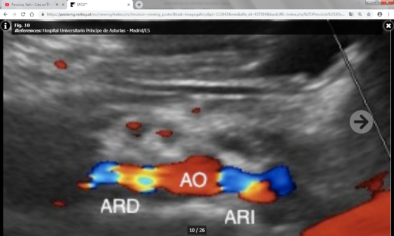

Ultrasound, the first-line imaging examination for RAS screening, commonly uses hemodynamic parameters for diagnosis ( 7), including peak systolic velocity (PSV), abdominal aortic velocity, the renal-aortic ratio (RAR) of the PSV and the presence of tardus-parvus renal artery spectral waveforms. Instead, contrast-enhanced ultrasound (CEUS), which uses contrast agents without hepatorenal toxic side effects, is especially suitable for patients allergic to ICAs or with renal insufficiency ( 6). In addition, iodinated contrast agents (ICAs) used in computed tomography angiography (CTA) and magnetic resonance angiography (MRA) carry the risk of contrast-induced nephropathy ( 4, 5). Digital subtraction angiography (DSA) is the gold standard for RAS diagnosis ( 2), but it is not a preferred imaging examination method due to its invasive nature and radiation exposure ( 3). Renal artery stenosis (RAS), which may lead to refractory hypertension and/or renal insufficiency, has been a substantial clinical concern ( 1).

RENAL ARTERY DOPPLER ULTRASOUND PROFESSIONAL
These all indicate the necessity and importance of the standardized operation of renal artery contrast-enhanced ultrasound examination, and professional training should be given to operators. The most important factor for the accurate diagnosis of renal artery stenosis by CEUS is the operator's standardized examination, which is not only related to the duration of the operator has been engaged in this inspection, but also related to whether the operator has received professional training in relevant aspects. CEUS combined with hemodynamic indicators (Doppler ultrasound) can improve the accurate diagnosis rate of renal artery stenosis by ultrasonography 4. CEUS showed good performance in detecting normal renal artery and renal artery stenosis, and the missed diagnosis is concentrated on mild and moderate stenosis 3. CEUS can clearly show the renal arteries, and is consistent with DSA in distinguishing normal renal artery stenosis from renal artery stenosis, as well as renal artery stenosis ≥70% and <70% stenosis 2. CEUS missed two cases (one for mild stenosis and one for moderate stenosis), and the detection rate of renal artery stenosis was 97.5% (78/80) A total of 65 renal arteries diagnosed by CEUS were consistent with DSA, and the diagnostic accuracy of CEUS for the degree of stenosis was 81.25% (65/80) Among the 13 misdiagnosed renal arteries, 4 of them can be corrected to the same degree as DSA by the reference to hemodynamic index, and the diagnosis rate of the degree of renal artery stenosis by ultrasonography (combined with CEUS and hemodynamic indicators) can be improved to 86.25%.Ĭonclusions: 1. Compared with the gold standard DSA results, the ROC curve of CEUS in distinguishing renal artery stenosis ≥70% from <70% stenosis has an AUC of 0.916, a sensitivity of 90.9%, a specificity of 92.3%, and the Kappa value of the consistency analysis is 0.77 ( P < 0.01) 3. Compared with the gold standard DSA results, the AUC of the ROC curve of CEUS in detecting normal renal artery and renal artery stenosis was 0.961, the sensitivity was 96.4%, the specificity was 95.8%, and the Kappa value of the consistency analysis was 0.912 ( P < 0.01) 2. Methods: The data of 40 patients (80 renal arteries) diagnosed with RAS by CEUS in Beijing Hospital from September 2018 to October 2020 were compared with their digital subtraction angiography (DSA) results to analyze the causes underlying missed diagnosis and misdiagnosis of RAS by CEUS. Objectives: To explore the role of Contrast-enhanced ultrasound (CEUS) in the evaluation of patients with suspected renal artery stenosis and analyze the causes of the misdiagnosis and missed diagnosis. Department of Ultrasound, Beijing Hospital, National Center of Gerontology, Institute of Geriatric Medicine, Chinese Academy of Medical Sciences, Beijing, China.Yang Wang, Yan Li, Siyu Wang, Na Ma and Junhong Ren *


 0 kommentar(er)
